
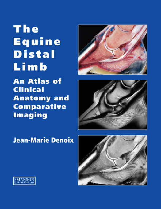
The Equine Distal Limb: Atlas of Clinical Anatomy and Comparative Imaging
Author/Editor: J.-M. Denoix
Publication Year: 2000
Overview
The Equine Distal Limb: Atlas of Clinical Anatomy and Comparative Imaging is an exceptionally detailed, visually rich reference correlating precise anatomical dissections with advanced diagnostic imaging. Authored by world-renowned equine anatomist and diagnostician J.-M. Denoix, this atlas bridges structural anatomy with radiography, ultrasonography, and magnetic resonance imaging. It stands as an indispensable manual for equine veterinarians, veterinary surgeons, and radiology specialists dedicated to precise lameness localization and diagnosis.
Key Topics Covered
Comprehensive Anatomical Dissections: Detailed photographic breakdowns of the equine foot, pastern, and fetlock across multiple spatial planes.
Comparative Sectional Anatomy: Sagittal, parasagittal, transverse, and frontal slice orientations designed to enhance three-dimensional comprehension.
Advanced Diagnostic Correlation: Integration of gross anatomical structures with modern imaging modalities to aid in accurate clinical interpretation.
Lameness & Pathology Insights: Targeted evaluations of complex distal limb structures to improve diagnostic accuracy and targeted treatment planning.



Reviews
There are no reviews yet.